Chronic nasal discharge or epistaxis cases can present a diagnostic challenge. A definitive diagnosis may require a combination of imaging, cytology, rhinoscopy, histopathology, and culture. Cytology from purulent nasal exudates often reveals septic inflammation which is consistent with rhinitis, but may be secondary to an underlying disease (fungal infection, foreign body, viral infection, or tumour). Deep swabs from the nasal cavity, especially if any mucosal plaques are observed, may reveal fungal hyphae in cases of fungal rhinitis.
Species:
All
Specimen:
Nasal discharge, aspirated material
Container:
Glass slides, slide container, exudate or aspirated fluid in a sterile container and EDTA, swab for culture
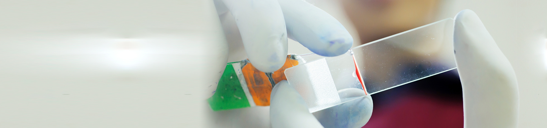
Collection protocol:
Slides: Place the nasal discharge on a glass slide. Then take another slide, gently place it on top. Applying a small amount of downward pressure to spread out the mucoid material, pull the slides along each other lengthwise until they separate. This will result in two slides with a monolayer of cells. If there is a detectable nasal mass, fine needle aspiration is recommended.
Aspirates: Submit fluid in both a plain container (for culture) and EDTA tube (for cytology).
Special handling/shipping requirements:
Place slides in slide-containers for shipment. Do not ship with samples containing formalin (wrap and bag separately). Formalin fumes prevent cellular uptake of differential stains, rendering the slides non-diagnostic. Do not store cytology slides in the fridge, as condensation will render the slides non-diagnostic.